基本介紹
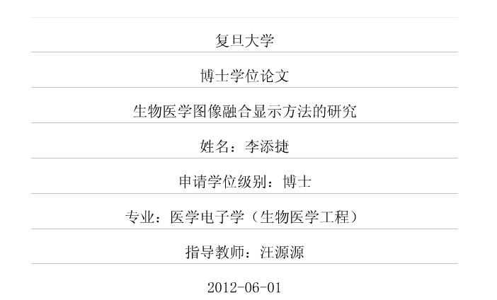
- 中文名:生物醫學圖像融合顯示方法的研究
- 類別:論文
- 摘要:成像技術層出不窮
- 關鍵字:廣義亮度-色相-飽和度框架
中文摘要
生物相襯圖與螢光圖的融合有助於蛋白質亞細胞結構的定位和功能研究。融合圖像應在真實反映細胞內蛋白質分布的基礎上,保留足夠的亞細胞結構的定位信息,著力改善螢光背景對融合圖像明度和清晰度的影響。
研究結合...>> 詳細
生物相襯圖與螢光圖的融合有助於蛋白質亞細胞結構的定位和功能研究。融合圖像應在真實反映細胞內蛋白質分布的基礎上,保留足夠的亞細胞結構的定位信息,著力改善螢光背景對融合圖像明度和清晰度的影響。
研究結合廣義亮度-色相-飽和度(Generalized Intensity-hue-saturation,GIHS)快速算法、非降採樣輪廓波變換(Nonsubsampled Contourlet Transform,NSCT)和透明度的概念,提出了GIHS框架下變權重多尺度融合的流程。融合方法的設計在引入了NSCT理論的基礎上,結合絕對值最大的高頻係數融合準則,提出了以雙伽馬函式為原型的低頻係數融合準則。
實驗部分結合Otsu分割算法和視覺信患保真度(Visual Information Fidelity, VIF)指標,設計了分區域的相似度量化實驗。通過117組擬南芥相襯圖和螢光圖的融合顯示實驗,研究了多尺度變換對圖像融合顯示效果的影響。實驗結果表明:非降採樣技術和二維濾波器設計在一定程度上提高了圖像融合的效果,但是多尺度變換的方向性與融合效果的提升關係不大。在該方法和多種傳統方法融合顯示效果的比較實驗中,量化結果與融合實例中的觀察結果吻合,都表明了該方法的有效性和相比傳統方法的優越性。
腦部磁共振成像(Magnetic Resonance Imaging,MRI)與單光子發射計算機斷層成像(Single Photon Emission Computed Tomography,SPECT)的融合實現了人體結構和功能信息的整合,尤其有助於SPECT圖的解讀和運用。
融合圖像應在真實反映腦部功能變化的基礎上,保留足夠的結構定位信息,著力於改善SPECT圖黑色背景對融合圖像明度和清晰度的影響。研究將圖像的融合顯示歸結為線性最佳化問題,運用單純形法得到權重函式,用於源圖像亮度分量低頻係數和高頻係數融合準則的設計。研究還設計了算法的可互動特性,提出了全局透明度和全局高頻透明度,用以實現源圖像之間的漸變以及對融合圖像細節表現的控制。
實驗探討了基於高階多項式近似的權重函式的估計,相比基於雙伽馬函式的近似,其優勢在於放寬了對權重函式形態的限制。在正常腦部29個層面MRI圖像和SPECT圖像的融合實驗中,融合實例的觀察結果與量化實驗結果吻合,都表明了該方法的有效性和相比傳統方法的優越性。繼而以傳統透明度技術為參照,驗證了融合顯示方法的可互動特性設計。兩組臨床實例證實了提出的融合顯示方法的實用價值。最後結合實驗,初步探討了兩種權重函式設計方法的優缺點和通用性。
B型超聲圖與超聲彈性圖的融合不僅有助於生物組織力學特性分布的定位、理解和運用,而且擴大了超聲無損診斷的套用範圍。融合圖像應在保持源圖像理解模式的基礎上,提高融合圖像對源圖信息的表現力,尤其是對B型超聲灰度圖像中低頻結構細節的表現力。
研究基於色貌模型理論,設計了均一明度的偽彩顯示方法,並將色貌屬性的預測用於圖像的融合顯示。在國際發光照明委員會CIECAM02色貌模型的運用中,通過假定已知明度、色相和飽和度,著重推導了圖像的CIEXYZ色彩空間表示。
實驗研究了計算機可顯示的色彩集合在色彩空間和色貌模型中的分布。其結果表明:灰度圖像的明度範圍受融合圖像中允許的色彩集合限制;色貌屬性中的飽和度調節了融合結果的色相解析度和明度範圍。通過49組仿真圖像的量化實驗,驗證了提出的基於CIECAM02色貌模型的融合顯示方法的有效性。結合2組臨床實例的融合結果,證實了該方法的套用價值,及其相比傳統方法的優越性。最後對套用範圍進行了擴展,初步探討了其在腦部MRI圖像融合中的可行性。
關鍵字:圖像融合顯示,廣義亮度-色相-飽和度框架、非降採樣輪廓波變換,變權重,伽馬函式,單純形法,色貌模型,生物醫學圖像
外文摘要
Combination of the fluorescent image and the phase contrast image is valuable for the localization and functional studies of the protein. The fused result aims to restrain the black background of the fluorescent image, and display a complete distribution of the targeted proteins with sufficient location information of subcelluar structures.
In the view of transparency, a variable-weight method is proposed in the generalized intensity-hue-saturation frame. The intensity components of original images are first analyzed by the nonsubsampled contourlet transform (NSCT). Subsequently, the low-frequency coefficients are combined as the rule based on a piecewise function consisting in two Gamma functions, while the high-frequency coefficients are combined as the maximum selection rule.
Using the Otsu method and the visual information fidelity (VIF), the region-based assessment of similarity is designed to reveal the similarity between the fused image and the original images. Validation experiments on 117 sets of Arabidopsis images are for two purposes: the impact of the multiscaled analysis in the fusion and the comparison among different methods of integrated visualization. It was demonstrated that the invariance and the design of true two-dimensional filters rather than the directionality attribute to the fused effect. Besides, both the quantitative results and the application examples demonstrated the superiority of the proposed method over the traditional ones.
The single photon emission computed tomography (SPECT) image alone is difficult to understand and utilize, since anatomical structures are absent from the data. Integrated visualization attempts to locate functional information of the SPECT image by the structural information of the magnetic resonance (MR) image. The fused result aims to restrain the black background of SPECT, and display a complete distribution of functional variation with sufficient location information of anatomical structures.
After approximating the weight by a polynomial function, integrated visualization can be regarded as a linear optimization problem in which the simplex method is applied to determine the coefficients of the weighting function. The rules are then proposed for the combination in the multiscaled spaces. Interactive approaches are designed for the gradual variation between original images with the global transparency, and the control of detail performance with the global high-frequency transparency.
In the experiments, the weighting function was first estimated. Its shape had less restraints than that based on two Gamma functions proposed in the former application. The similarity assessment then evaluated several different methods on a normal brain atlas composed of 29 slices. The observations in the example matched the quantitative results, and demonstrated the superiority of the proposed method over the traditional methods. Two clips showed the interactive property of the proposed method, while two medical cases demonstrated its clinical values. Besides, two methods for designing the S-shaped weighting function were also compared, ie. the simplex method and the method based on two Gamma functions.
Combined with the B-mode ultrasound image, the elastography image can be better understood and utilized, which also expands the application range of ultrasonic non-destructive examination. Except preserving the original styles of interpretation, the fused result also aims to improve the ability in expressing the original images, especially the low-frequency structures in the B-mode ultrasound image.
The fused method is proposed based on the color prediction of the color appearance model. It additionally involves the pseudo-color display with uniform lightness. Here the formulae are exhaustively derived from the CIECAM02 color attributes to the CIEXYZ color channels, after the assumption of the known attributes including lightness, hue and saturation.
Based on different color spaces and the color appearance models, the distribution of color aggregations was studied in the experiment. It was concluded that the range of lightness was determined by the available color aggregation for the fused image, and the saturation controlled the resolution of hue and the range of lightness. The similarity assessment was performed on 49 sets of simulated images. Its result was reasonable, and thus revealed the effectiveness of the proposed method. Two medical cases demonstrated the clinical values of the proposed method and its superiority over the traditional methods. Besides, its feasibility in fusing two high-resolution structural images was preliminarily approved based on the simulated MRI data.
Keywords: integrated visualization, generalized intensity-hue-saturation (GIHS), nonsubsampled contourlet transform

